IRON DEFICIENCY ANEMIA IN ADULTS
"Anemia is a late finding due to iron deficiency."
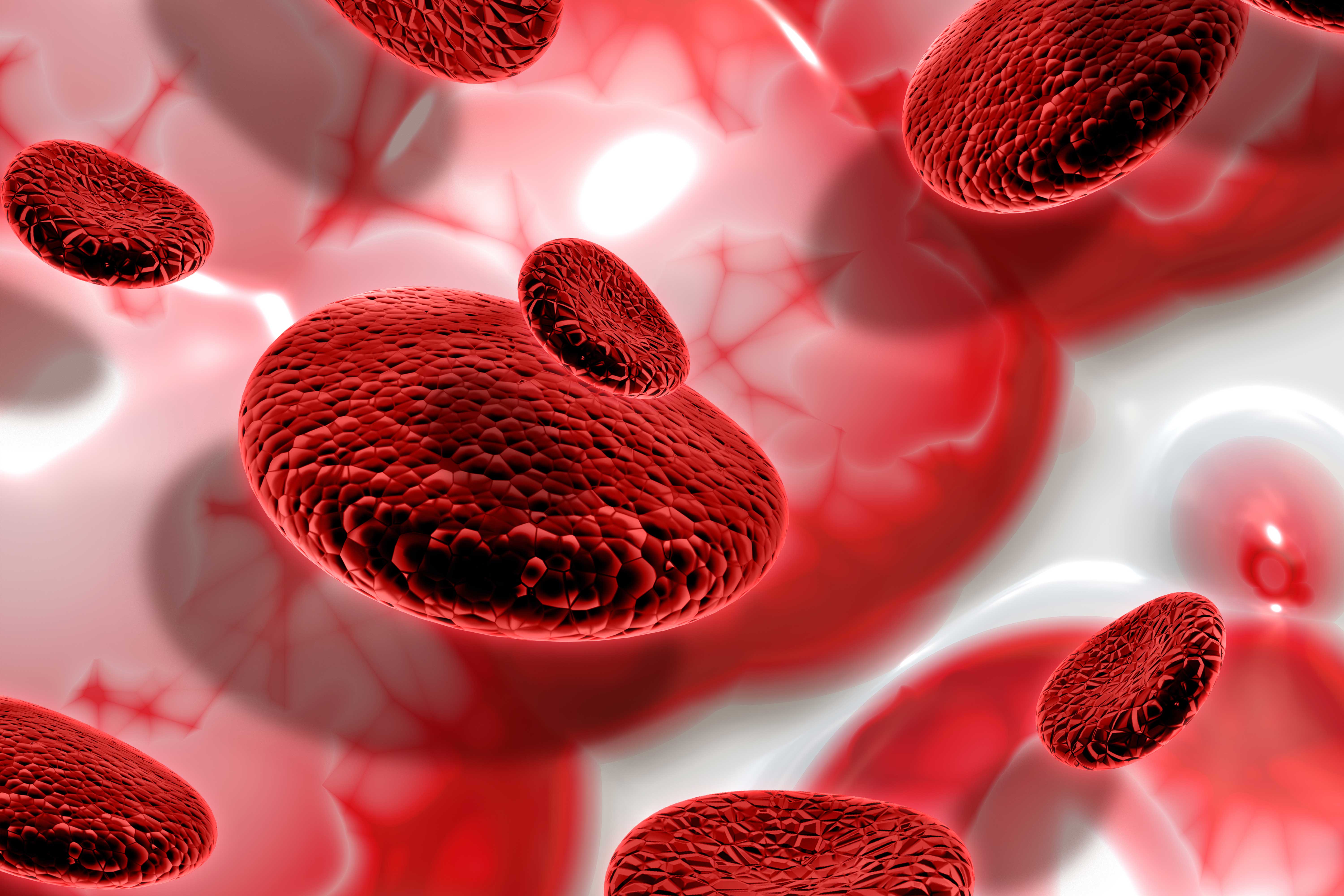
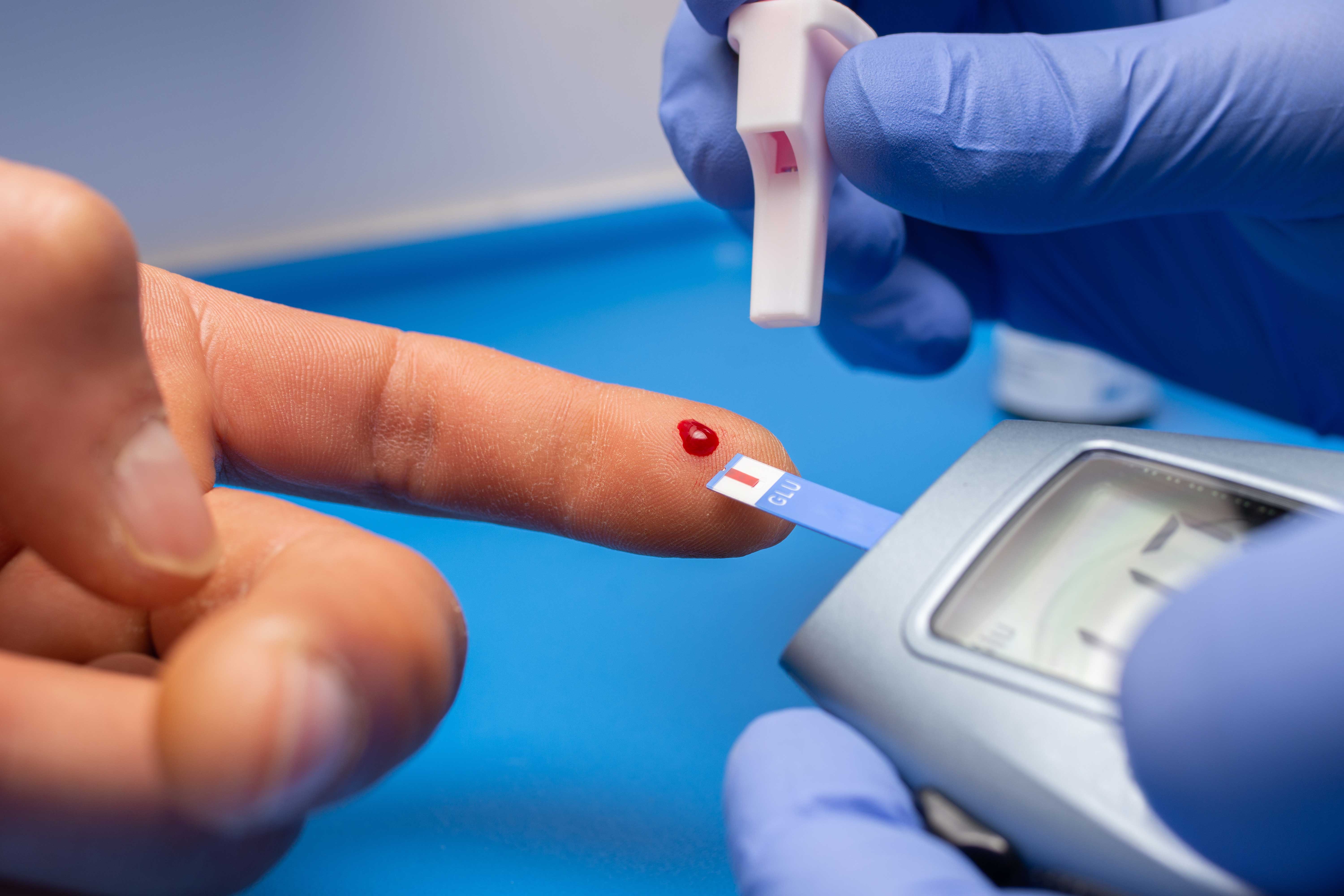
What is anemia?
Causes and risk factors of iron deficiency anemia
When we look at the causes of iron deficiency anemia, we can count three main causes; inadequate intake of iron from food, conditions with iron malabsorption (imperfect absorption), and iron loss more than iron intake.
In adults in developed countries, dietary intake is usually adequate and it is important to assume that the cause is blood loss until proven otherwise and to investigate this as a priority. However, this is not the case for underprivileged populations. As in the diagnosis of many diseases, a good history is the most important factor in determining the cause of iron deficiency anemia. Nutritional status, the presence of diarrhea in terms of possible malabsorption, previous gastrointestinal surgery, medications, and the presence of any bleeding foci should be questioned in detail.
2.1. Reduced iron absorption
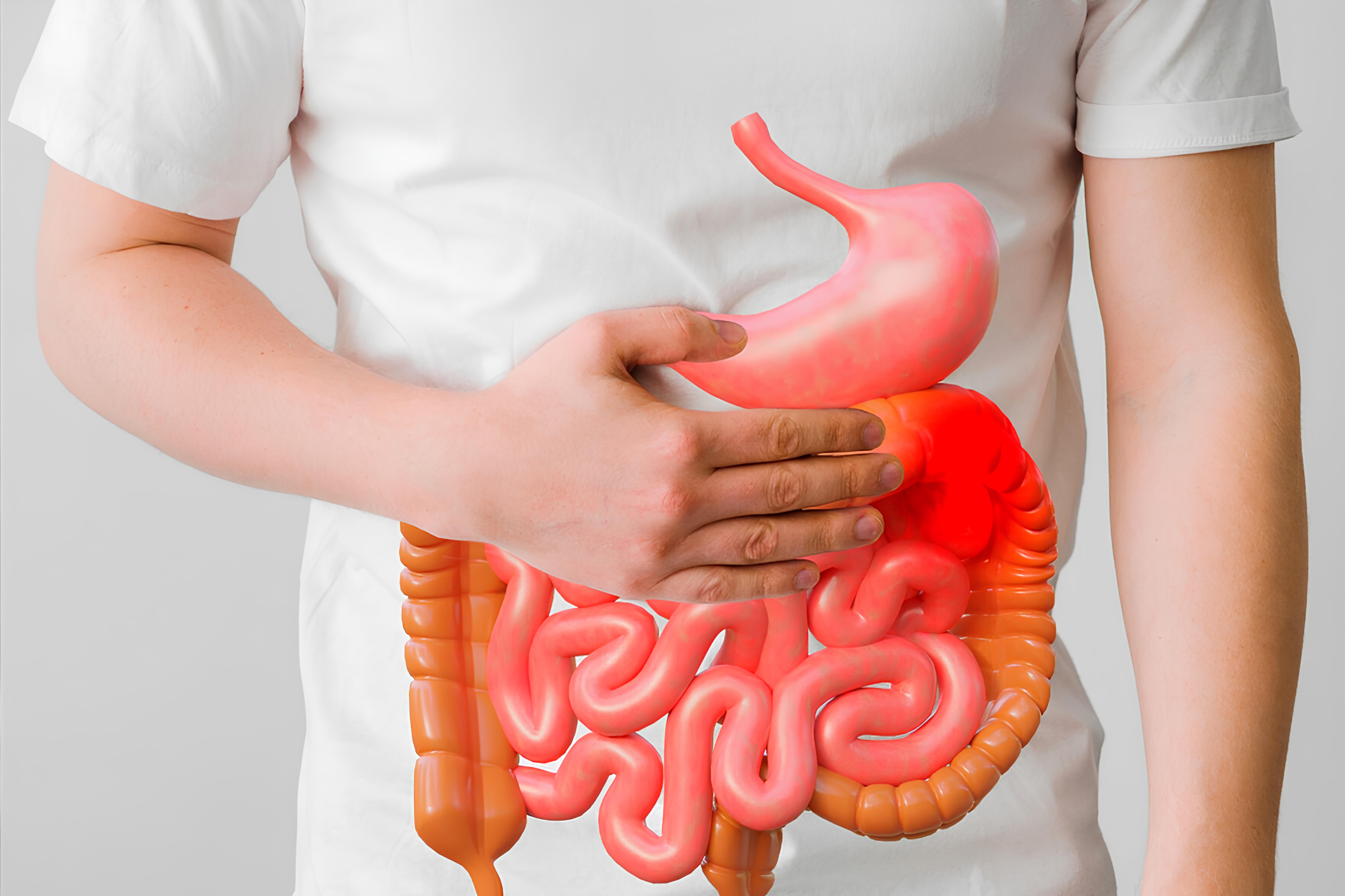
Iron is absorbed in the upper gastrointestinal tract, with the duodenum being the site of highest absorption [3]. Reduced iron absorption is a rare cause of iron deficiency, especially in healthy individuals and in parts of the world where there is access to an iron-rich diet.
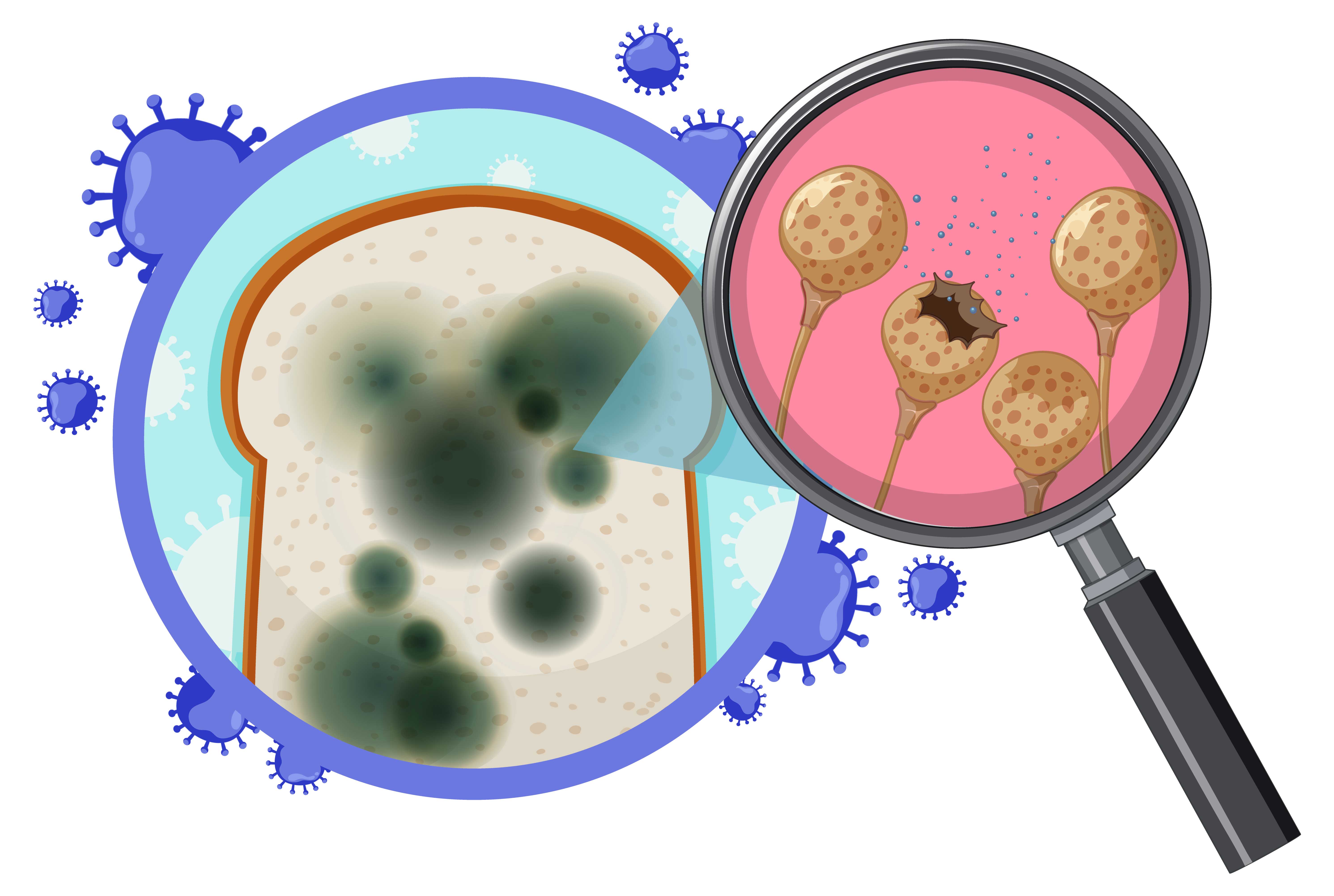
Disorders affecting mucosal cells responsible for iron absorption such as celiac disease, atrophic (autoimmune*) gastritis, Helicobacter pylori infection, and bariatric surgery (surgery for obesity that cannot be treated with diet and exercise) are important causes of malabsorption (imperfect absorption).
In people with gastrointestinal symptoms or inadequate response to oral iron supplementation, sources of reduced iron absorption may be considered. A source of bleeding should be excluded, whether or not reduced iron absorption has been documented.
*Autoimmune: A situation in which a person creates an immune response against his or her own cells and tissues as a result of a disorder in the immune system.
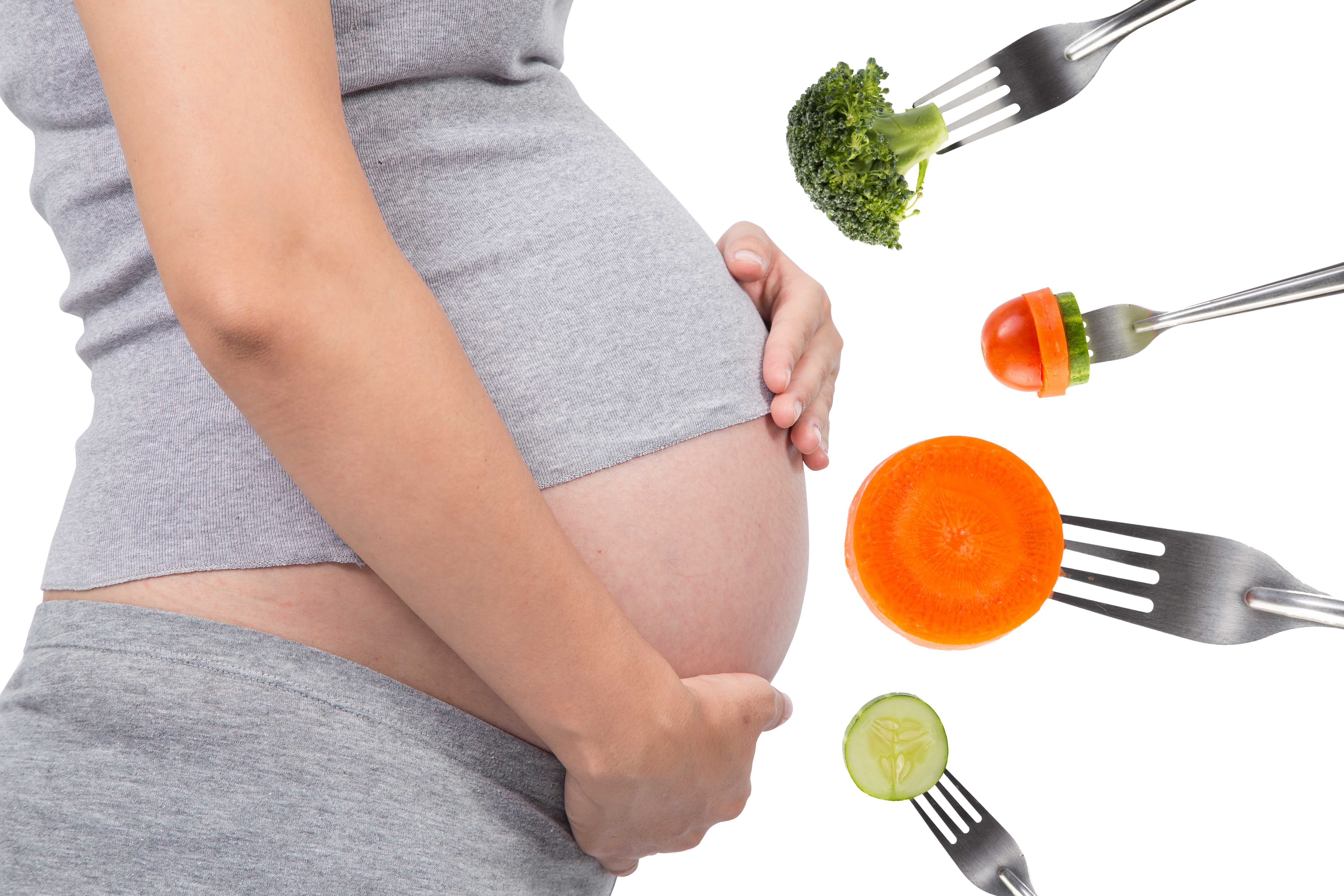
2.2. Dietary
Heme iron (iron from meat rather than plant sources) is better absorbed than non-heme iron, but both sources are important. A review of published studies assessing vegetarians and non-vegetarians found that those who consumed a vegetarian diet were more likely to develop iron deficiency [4].
Some foods can impair iron absorption, such as tannins (found in tea), phosphates, phytates (mineral-binding compounds found in whole grains and seeds), and foods high in calcium
One case report described iron deficiency in an individual who drank excessive amounts of tea (more than 1500 mL per day for 20 years). Upper and lower endoscopies showed no pathological conditions and hemoglobin increased with cessation of tea drinking and decreased with continued tea drinking [5].
2.3. Celiac disease (gluten-sensitive enteropathy)
Celiac disease (gluten-sensitive enteropathy) is a small intestinal malabsorption disorder triggered by exposure to gluten in susceptible individuals. Iron may contribute to anemia through several mechanisms, including malabsorption (imperfect absorption) of other nutrients required for red blood cell (RBC) production, including vitamin B12, folic acid, and copper [6].
2.4. Autoimmune gastritis or Helicobacter pylori
Autoimmune gastritis or Helicobacter pylori (H. pylori); gastritis due to an autoimmune mechanism (e.g., anti-parietal cell antibodies) or H. pylori has also been shown to be an important cause of iron deficiency [7,8].
2.5. Bariatric surgery
Iron requires to be conjugated with vitamin C, amino acids, or sugars in the presence of gastric acid to protect it from alkaline (in a base pH) secretions in the proximal jejunum; otherwise, iron is converted to ferric hydroxide and cannot be absorbed. Bariatric surgery involves a number of procedures that promote weight loss by limiting the reservoir capacity of the stomach and/or shortening the length of the functional small intestine, leading to malabsorption (imperfect absorption). The risk of causing iron deficiency is highest as it reduces the absorption area. Monitoring iron status is recommended after most bariatric surgeries with routine iron supplementation or iron supplementation as needed.
2.6. Drugs
An acidic gastric environment facilitates the absorption of iron, especially non-heme iron. Reduced gastric acidity alone is unlikely to cause clinically significant iron deficiency in an individual with adequate dietary iron intake and a normally functioning gastrointestinal tract.
Drugs that reduce gastric acidity, especially proton pump inhibitors (PPIs, stomach protector), have been suggested to decrease iron absorption [9,10].
2.7. Inherited disorders that interfere with iron absorption
Inherited disorders that interfere with iron absorption are very rare. IRIDA (iron therapy-resistant iron deficiency anemia) due to a TMPRSS6 mutation is a rare inherited disorder in which oral iron absorption is markedly impaired. IRIDA is caused by loss-of-function mutations of the TMPRSS6/matriptase 2 gene, which encodes a serine protease that cleaves membrane-bound hemojuvelin [11,20]. It causes iron deficiency due to inappropriately high levels of hepcidin, markedly decreased iron absorption, and increased iron uptake in macrophages* [17, 21-26]. Patients present with mild hypochromic*, microcytic anemia*** with very low serum iron levels and low transferrin saturation. Serum ferritin levels are usually in the normal range or slightly elevated following intravenous iron therapy [17].
*Macrophage: These are white blood cells that protect the body against diseases.
**Hypochromia: The amount of hemoglobin in the red blood cell is below the reference range of 32 - 36 grams/deciliter (g/dL).
***Microcytic anemia: It suggests a type of anemia in which red blood cells are smaller than normal.
2.8. Iron deficiency anemia due to iron loss
Menstrual bleeding is the most important cause of iron deficiency anemia in premenopausal women. Gastrointestinal bleeding in postmenopausal women and adult men is an important cause of IDA. Further investigations are absolutely necessary. Upper and lower gastrointestinal endoscopy is recommended to rule out the bleeding focus and possible malignancy (types of tumors that can be considered cancer). Bleeding disorders such as Von Willebrand Disease should also be kept in mind, especially in prolonged menstrual bleeding.
Symptoms of anemia
Symptoms of anemia
Treatment
1. Ferhanoğlu
B. PDQ Hematoloji (William F.Kern, MD). 1. Baskı, İstanbul Medikal Yayıncılık,
2005: 1-155
2. Ünal
S. Cecil Texbook of Medicine (L. Goldman, D. Ausiello). 22. Baskı. Güneş
Yayınevi, 2006:963-75
3. Schröder
O, Mickisch O, Seidler U, ve diğerleri. İnflamatuar barsak hastalığı olan
hastalarda demir eksikliği anemisinin tedavisinde intravenöz demir sükroza
karşı oral demir takviyesi - randomize, kontrollü, açık etiketli, çok merkezli
bir çalışma. Am J Gastroenterol 2005; 100:2503.
4. Gasche
C, Berstad A, Befrits R, ve diğerleri. İnflamatuar barsak hastalıklarında demir
eksikliği ve aneminin tanı ve tedavisine ilişkin kılavuzlar. Inflamm Bowel Dis
2007; 13:1545.
5. Reinisch
W, Chowers Y, Danese S, ve diğerleri. İnflamatuar barsak hastalığında demir
eksikliğinin yönetimi – RAND/UCLA uygunluk yöntemiyle geliştirilen çevrimiçi
bir araç. Aliment Pharmacol Ther 2013; 38:1109.
6. Gasche
C, Evstatiev R, Haas T, ve diğerleri. İnflamatuar barsak hastalıklarında demir
eksikliği ve aneminin tanı ve tedavisi. Avusturya IBD Çalışma Grubunun
Mutabakatı]. Z Gastroenterol 2011; 49:627.
7. Mamula
P, Piccoli DA, Peck SN, ve diğerleri. İnflamatuar barsak hastalığı olan
çocuklarda demir eksikliği anemisi için toplam doz intravenöz demir dekstran
infüzyonu. J Pediatr Gastroenterol Nutr 2002; 34:286.
8. Lindgren
S, Wikman O, Befrits R, ve diğerleri. İBH hastalarında aneminin düzeltilmesi ve
demir depolarının yenilenmesi açısından intravenöz demir sükroz, oral demir
sülfattan daha üstündür: Randomize, kontrollü, değerlendiriciden kör, çok
merkezli bir çalışma. Scand J Gastroenterol 2009; 44:838.
9. Kim
YW, Bae JM, Park YK ve diğerleri. Gastrektomi Sonrası Akut İzovolemik Anemili
Hastalarda İntravenöz Ferrik Karboksimaltozun Hemoglobin Yanıtı Üzerine Etkisi:
FAIRY Randomize Klinik Çalışması. JAMA 2017; 317:2097.
10. Auerbach
M, Achebe MM, Thomsen LL, Derman RJ. Bariatrik cerrahi sonrası demir eksikliği
anemisi olan hastalarda demir sükroz (IS) ile karşılaştırıldığında ferrik
derozomaltozun (FDI) etkinliği ve güvenliği. Obes Cerrahisi 2022; 32:810.
11. Khalafallah
AA, Yan C, Al-Badri R, ve diğerleri. Postoperatif anemi tedavisinde intravenöz
ferrik karboksimaltoza karşı standart bakım: prospektif, açık etiketli,
randomize kontrollü bir çalışma. Lancet Haematol 2016; 3:e415.
12. Hershko
C, Ronson A. Demir eksikliği, Helicobacter enfeksiyonu ve gastrit. Acta
Haematol 2009; 122:97.
13. Auerbach
M, Liang AS, Glaspy J. Kemoterapide ve kansere bağlı anemide intravenöz demir.
Topluluk Oncol 2012; 9:289.
14. Gafter-Gvili
A, Rozen-Zvi B, Vidal L, ve diğerleri. Kemoterapinin neden olduğu aneminin
tedavisi için intravenöz demir takviyesi - randomize kontrollü çalışmaların
sistematik incelemesi ve meta-analizi. Acta Oncol 2013; 52:18.
15. Rodgers
GM 3rd, Becker PS, Blinder M, ve diğerleri. Kanser ve kemoterapinin neden
olduğu anemi. J Natl Compr Canc Netw 2012; 10:628.
16. Kim
YT, Kim SW, Yoon BS ve diğerleri. Eş zamanlı kemoradyoterapi ile tedavi edilen
serviks kanseri hastalarında intravenöz olarak uygulanan demir sükrozun aneminin
önlenmesine etkisi. Gynecol Oncol 2007; 105:199.
17. Dangsuwan
P, Manchana T. Kemoterapi alan jinekolojik kanser hastalarında intravenöz demir
ile kan transfüzyonunun azaltılması. Gynecol Oncol 2010; 116:522.
18. Steinmetz
T, Tschechne B, Harlin O, ve diğerleri. Kanser ve kemoterapiye bağlı anemi
tedavisinde ferrik karboksimaltoz ile klinik deneyim. Ann Oncol 2013; 24:475.
19. Spivak
JL. Polisitemi vera: mitler, mekanizmalar ve yönetim. Kan 2002; 100:4272.
20. Clénin
G, Cordes M, Huber A, ve diğerleri. Sporda demir eksikliği - tanımı, performans
ve tedaviye etkisi. İsviçre Med Haftalık 2015; 145:w14196.
21. Alaunyte
I, Stojceska V, Plunkett A. Iron ve kadın sporcu: demir durumunu ve egzersiz
performansını iyileştirmeye yönelik diyet tedavisi yöntemlerinin gözden
geçirilmesi. J Int Soc Spor Nutr 2015; 12:38.
22. Tolkien
Z, Stecher L, Mander AP ve diğerleri. Demir sülfat takviyesi yetişkinlerde
önemli gastrointestinal yan etkilere neden olur: sistematik bir inceleme ve
meta-analiz. PLoS One 2015; 10:e0117383.
23. Bregman
DB, Morris D, Koch TA, ve diğerleri. Hepsidin düzeyleri, demir eksikliği
anemisi olan hastalarda oral demir tedavisine yanıtsızlığı öngörmektedir. Am J
Hematol 2013; 88:97.
24. Werner
E, Kaltwasser JP, Ihm P. [Ağızdan demir tedavisi: bağırsak emilimi ve bir
yemeğin etkisi (yazarın çevirisi)]. Dtsch Med Wochenschr 1977; 102:1061.
25. Bazeley
JW, Wish JB. KBH Anemisinde Demir Eksikliği için Yeni ve Gelişen Tedaviler: Bir
Gözden Geçirme. Am J Böbrek Dis 2022; 79:868.
26. Powers
JM, Buchanan GR, Adix L, ve diğerleri. Beslenmeyle İlgili Demir Eksikliği
Anemisi Olan Küçük Çocuklarda Düşük Doz Demir Sülfat ve Demir Polisakkarit
Kompleksinin Hemoglobin Konsantrasyonu Üzerine Etkisi: Randomize Bir Klinik
Çalışma. JAMA 2017; 317:2297.
27. Lee
T, Clavel T, Smirnov K, ve diğerleri. Oral ve intravenöz demir replasman
tedavisi, İBH hastalarında bağırsak mikrobiyotasını ve metabolomunu belirgin
şekilde değiştirir. Gut 2017; 66:863.
28. Cook
JD, Reddy MB. Askorbik asit alımının tam bir diyetten hem içermeyen demir
emilimine etkisi. Am J Clin Nutr 2001; 73:93.
29. Cancelo-Hidalgo
MJ, Castelo-Branco C, Palacios S, ve diğerleri. Farklı oral demir
takviyelerinin tolere edilebilirliği: sistematik bir inceleme. Curr Med Res
Görüşü 2013; 29:291.
30. Macdougall
IC, Strauss WE, McLaughlin J, ve diğerleri. KBH'li hastalarda demir eksikliği
anemisinin tedavisinde ferumoksitol ve demir sükrozun randomize bir
karşılaştırması. Clin J Am Soc Nephrol 2014; 9:705.7.


Hypertension

Biomaterials in Neural Tissue Engineering

Pregnant Health

Child Health

Probiotics
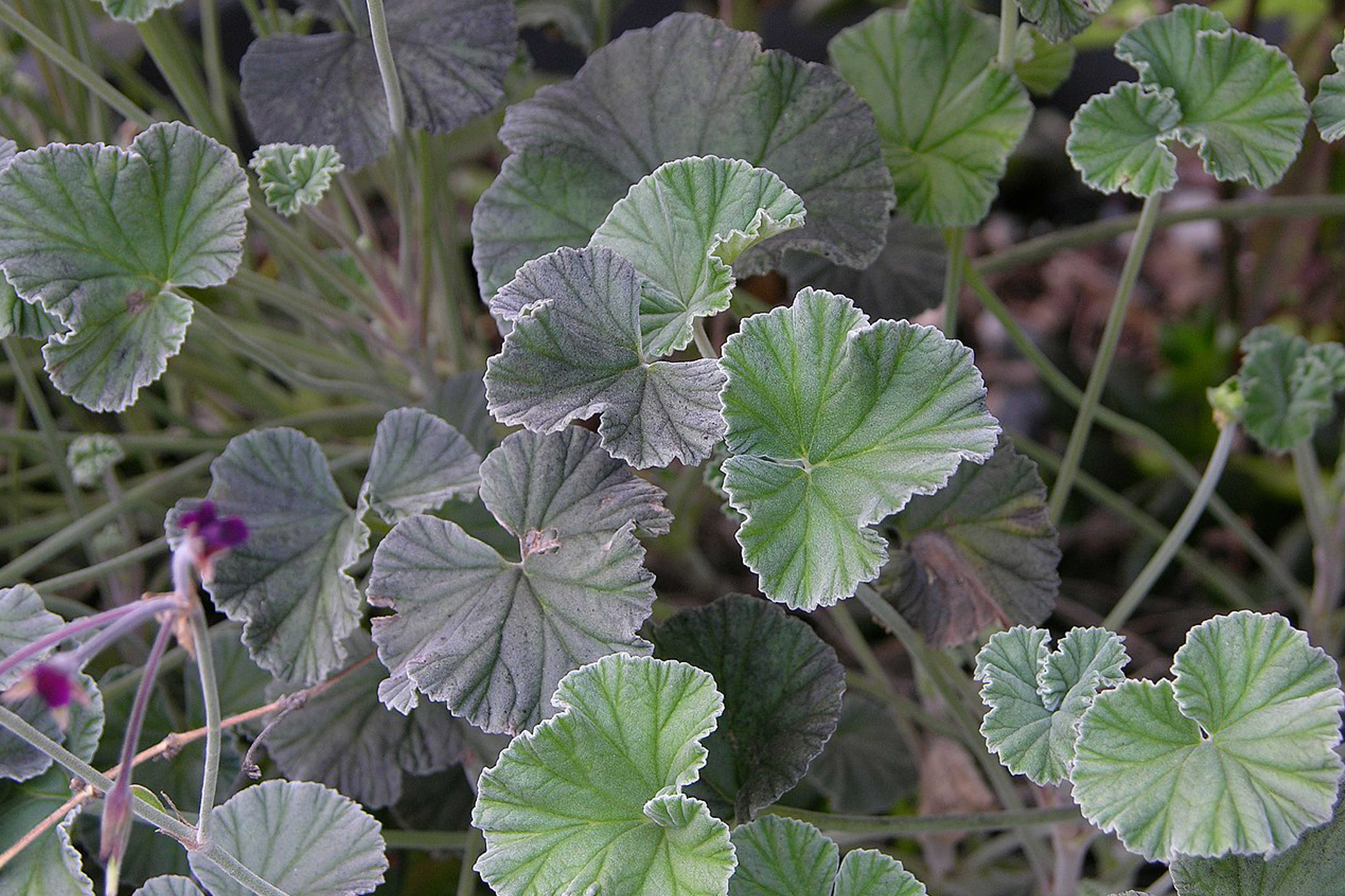
Afrikan Geranium
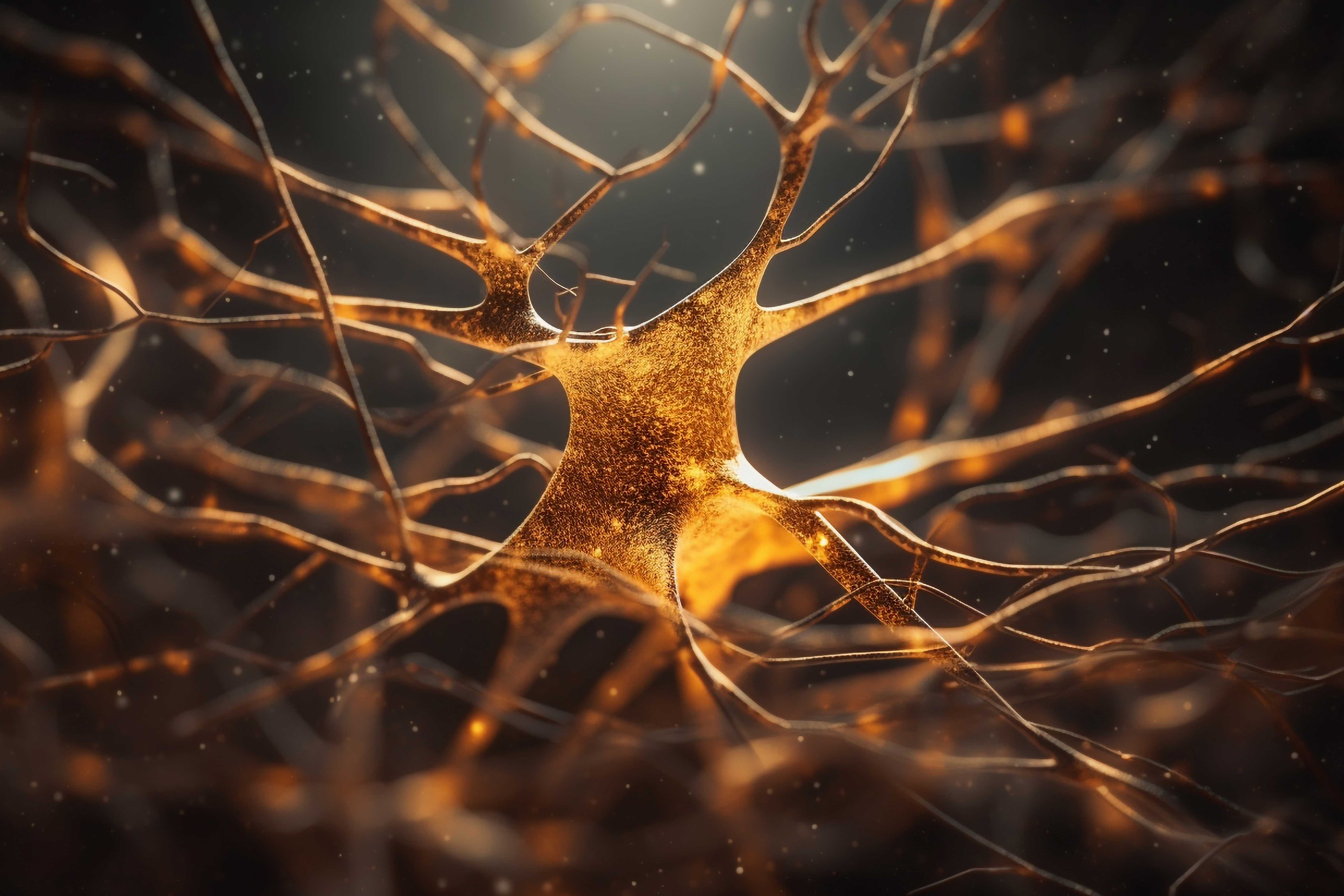
Biomaterials in Neural Tissue Engineering

BIRTH OF ARTIFICIAL INTELLIGENCE

Everything You Need To Know About Epilepsy
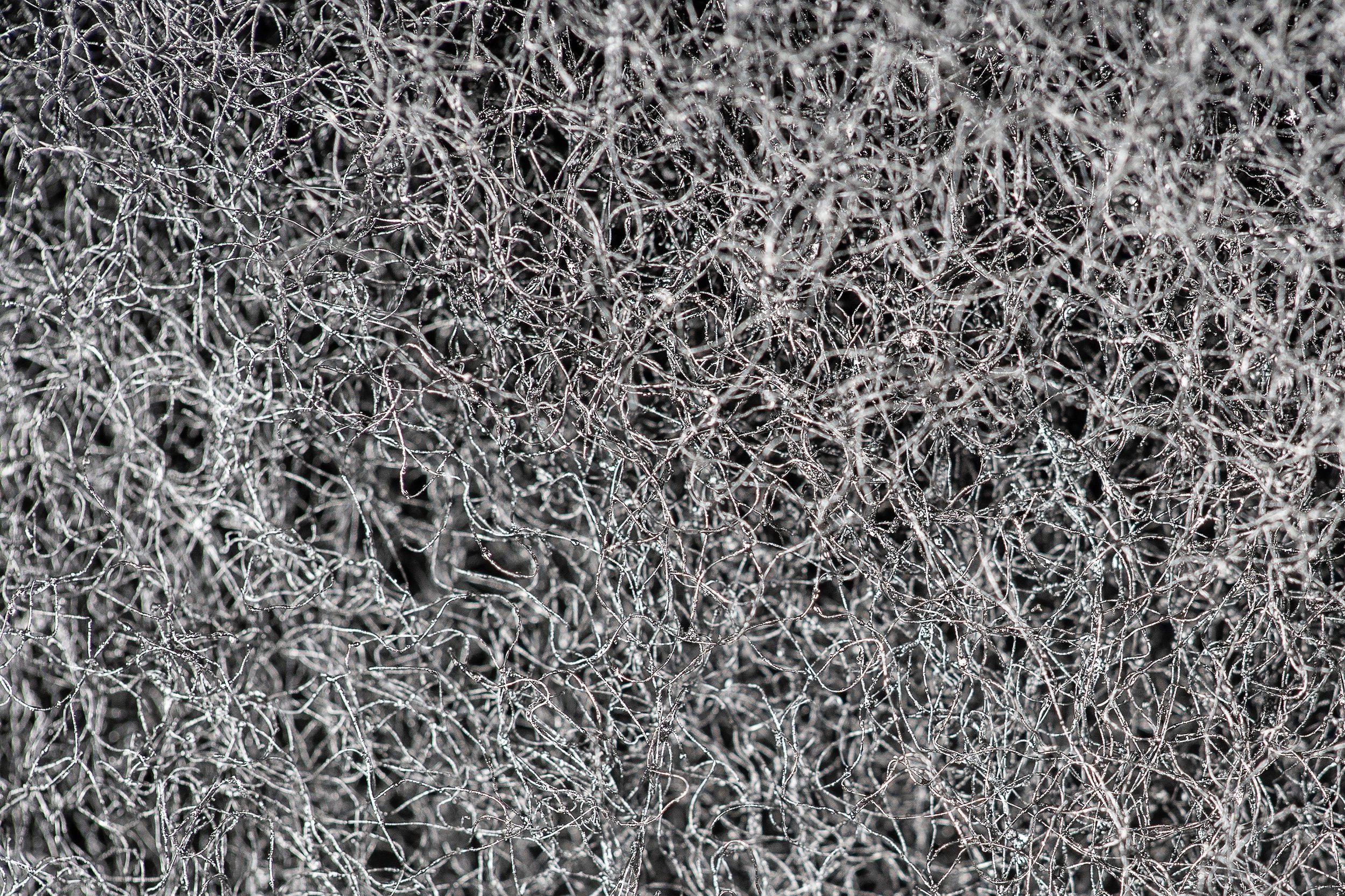
NANOFIBRES

Microneedles

3-Dimensional (3D) Bioprinting Technology and Bioprinters

REFLECTIONS OF DIGITAL APPLICATIONS IN PHARMACY COMMUNICATION ON MEDICINE AND HEALTH SERVICES

INSOMNIA

A Look At Headaches As The Crow Flies

HAIR PROBLEMS AND SOLUTIONS
Pharmacological Importance of Agmatine in the Brain

HIV/AIDS

Cough

CHRONIC STRESS AND FATIGUE: IN-DEPTH ANALYSIS

Blepharitis: What is it and how is it treated?
EVERYTHING YOU NEED TO KNOW ABOUT EPILEPSY

Depressive Disorders

Aromatherapy
Oral and Dental Health

Psoriasis from All Aspects

Beyond The Scale: Understanding The Complexities of Obesity

GENERAL INFORMATION ABOUT CANCER
IRON DEFICIENCY ANEMIA IN ADULTS
EVERYTHING YOU NEED TO KNOW ABOUT HEART FAILURE
Diabetes Mellitus
Crohn s Disease

The World's Longest-Acting Insulin Candidate in Diabetes Treatment: Insulin Icodec

Big Pharma Skepticism – What are we consuming?

Skin Health

Seborrheic Dermatitis

Acne Vulgaris

SKIN AGING AND TEA
NUTRITION IN THE PRECONCEPTİON PERIOD

Obesity

POPULAR DIET PERCEPTION

FUNCTIONAL FOODS IN WEIGHT MANAGEMENT
AFLATOXINS, THE HIDDEN POISON IN FOOD

A POWERFUL ALLY IN THE FIGHT AGAINST INFECTIOUS DISEASES: NUTRITION
